MRI & Advanced Diagnostic Imaging In Elmhurst IL
MRI & Other Diagnostics are crucial medical tools that offer life-saving solutions for countless patients globally. These diagnostics have transformed the medical field by enabling doctors to diagnose medical conditions more accurately and efficiently. Find out how various aspects of MRI and other diagnostic technologies have transformed medical diagnosis.
The Importance of Diagnostic Imaging Techniques in Orthopaedics

Regarding orthopedics, accurate diagnosis, and treatment planning is crucial for a successful outcome. Diagnostic imaging techniques play a critical role in helping to evaluate the complex system of bones, joints, muscles, tendons, and ligaments. At Orthopedic Specialists, we recognize the significance of these procedures, which aid in finding the source of pain and discomfort, analyzing the severity of the injury or ailment, and designing the most suitable course of therapy, all while being directed by our specialists.
Magnetic Resonance Imaging (MRI) Scan

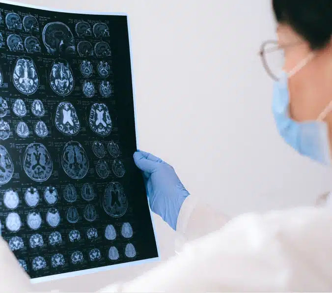
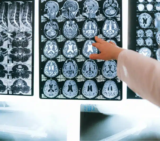
MRI scans use a powerful magnetic field and radio waves to create detailed body images. They are particularly useful in visualizing soft tissues, such as cartilage and ligaments, making them ideal for diagnosing conditions such as torn ligaments and herniated discs. MRI scans do not use radiation, making them a safe alternative to other imaging techniques. However, they are more expensive than X-rays, and CT scans, and not all patients are suitable candidates for MRI imaging.


Computed Tomography (CT) Scan

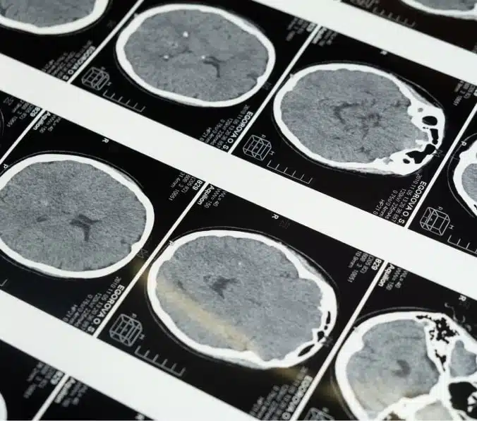
CT scans use X-rays to create a 3D image of the body. They are especially useful in identifying complex fractures, such as those in the spine or pelvic bones. CT scans also effectively image soft tissues, such as muscles and tendons. However, because of their higher radiation dose than X-rays, frequent CT imaging can increase cancer risk, especially in young patients.
X-Rays (Radiographs)

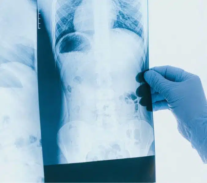
X-rays are the oldest and most widely used diagnostic imaging technique in orthopedics. An X-ray is a form of electromagnetic radiation that passes through the body and is detected on film or digitally. X-rays are beneficial for identifying bone fractures, dislocations, and osteoporosis. They can also detect calcifications, such as those seen in some types of arthritis. However, X-rays are limited in showing soft tissues, such as tendons and ligaments, making them less effective in diagnosing certain orthopedic conditions, such as rotator cuff injuries.


Other Orthopaedic Imaging Studies

Other imaging techniques, such as ultrasound and bone scan, are also used in orthopedic diagnosis. Ultrasound uses high-frequency sound waves to create images of the body and is particularly useful in diagnosing conditions of the tendons and muscles. A bone scan uses a small amount of radioactive material to identify areas of increased bone activity, such as those seen in cancer or infection. These techniques are generally safer than other imaging techniques but are less commonly used in orthopedics than X-rays, CT scans, and MRI scans.
Why Choose Orthopedic Specialists
At Orthopedic Specialists, we use state-of-the-art diagnostic imaging techniques as they play a critical role in diagnosing and treating orthopedic conditions. X-rays are the go-to imaging technique for diagnosing bone fractures and osteoporosis. At the same time, CT and MRI scans effectively diagnose complex fractures and soft tissue injuries. Other imaging techniques, such as ultrasound and bone scan, are helpful in certain situations. Patients should always consult with their orthopaedist before undergoing any imaging study, as each imaging technique has its benefits and limitations.
Book Your AppointmentFAQs
In orthopedics, an MRI is used for detailed imaging of bones, joints, muscles, tendons, and ligaments. It assists in diagnosing various conditions, including fractures, arthritis, cartilage damage, and soft tissue injuries, while helping plan appropriate treatments and monitor progress.
An orthopedic CT scan is a specialized imaging technique to visualize bones, joints, and surrounding structures in high-resolution detail. This non-invasive procedure employs X-ray technology to create cross-sectional images, aiding in diagnosing fractures, bone abnormalities, arthritis, and other musculoskeletal conditions while guiding treatment plans and surgical procedures.
An MRI scan utilizes magnetic fields and radio waves to produce detailed images of soft tissues. In contrast, a CT scan employs X-rays for high-resolution images of bones and dense tissues. Both provide valuable diagnostic information but cater to different clinical needs.
You may need an MRI scan when experiencing persistent pain, swelling, or limited mobility, requiring a detailed evaluation of soft tissues, bones, and joints. Physicians often recommend MRI scans for diagnosing injuries, monitoring treatment progress, or assessing the severity of certain conditions.
MRIs and CT scans are important as they provide detailed internal images, aiding in accurate diagnosis and treatment planning. MRIs excel at visualizing soft tissues, while CT scans focus on bones and dense structures. These imaging techniques enable physicians to make informed decisions, improving patient outcomes and recovery.
MRI scans are generally safe as they use non-ionizing radiation, unlike X-rays or CT scans. However, specific contraindications, such as metal implants, pacemakers, or pregnancy, may require extra precautions. Always inform your healthcare provider of any concerns or pre-existing conditions before undergoing an MRI.
An MRI with contrast involves using a contrast agent, typically gadolinium-based, injected into the patient’s bloodstream during the scan. This enhances the visibility of specific tissues and structures, allowing for improved image clarity and detail and assisting in a more accurate diagnosis and assessment of certain conditions.